
Updated on February 20, 2024
Corneal Pachymetry: Measuring Corneal Thickness


Vision Center is funded by our readers. We may earn commissions if you purchase something via one of our links.
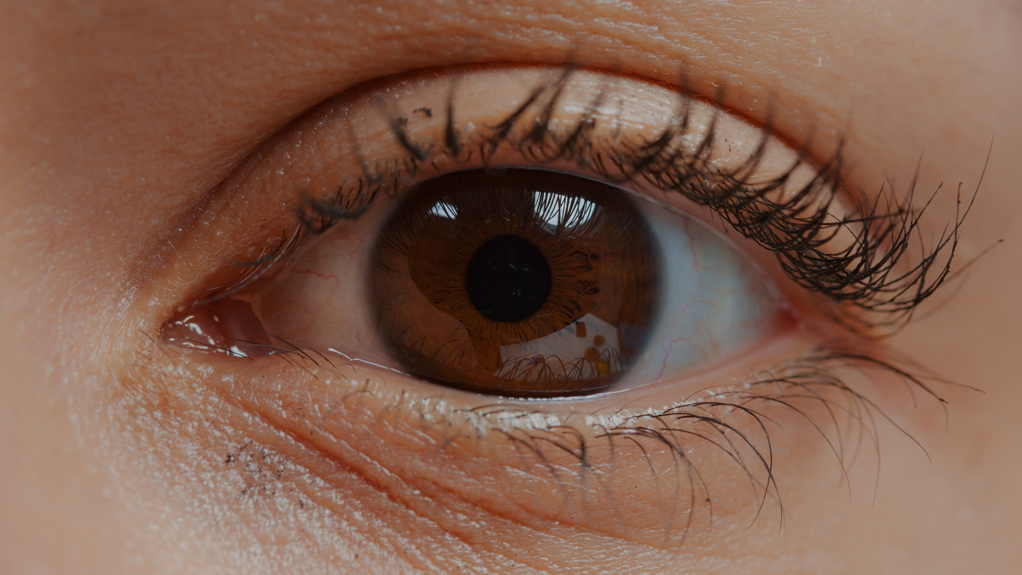
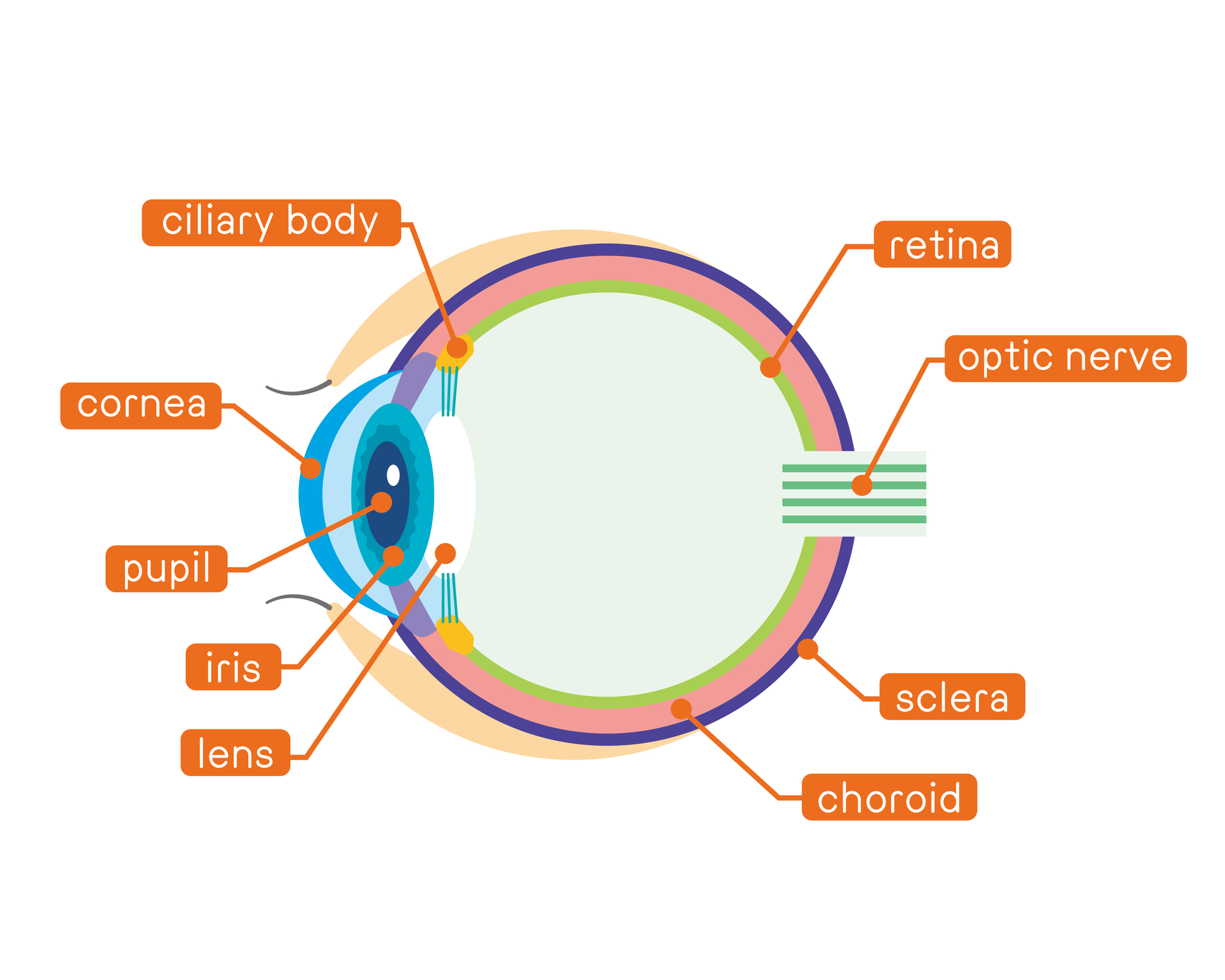
Corneal pachymetry is an exam to measure the cornea’s thickness. Eye doctors do this by using a medical instrument called a pachymeter. Pachymetry is a quick and painless test that has many uses.
Your eye doctor may perform corneal pachymetry to diagnose and monitor certain diseases, including glaucoma. Pachymetry also allows eye surgeons to determine eligibility for LASIK, which helps avoid potential complications.
This article explains the importance of taking corneal thickness measurements and how corneal pachymetry is performed.
Why Is it Important to Measure Corneal Thickness?
Pachymetry is a simple test, but it plays an important role in clinical ophthalmology. Here are some common reasons your doctor might take a corneal thickness measurement:
Determining the Presence of Corneal Swelling
Pachymetry will tell your doctor if you have any inflammation or swelling in your cornea. This helps your doctor diagnose certain diseases and issues.
Fuch's Dystrophy is a disease that causes fluid buildup in the cornea, which makes it swell. This increases the thickness of the cornea. Wearing contact lenses can also cause the cornea to swell.
Increases in corneal thickness can be difficult to detect with a microscope, which is why pachymetry is helpful.
Glaucoma Testing
More recently, pachymetry has become important in glaucoma investigation. Glaucoma is characterized by increased eye pressure, also known as intraocular pressure.
Studies show a close relationship between central corneal thickness and the risk of developing glaucoma.3 Increased intraocular pressure in the eyes can damage your optic nerve fibers. This can result in primary open-angle glaucoma or vision loss.4
Monitoring Glaucoma Development
People who have glaucoma need their intraocular pressure measured to monitor the condition. Corneal thickness affects the overall pressure measurement.
People with thin corneas (less than 540 microns) have a higher chance of low eye pressure readings even when the pressure is significantly high.7 This is dangerous because high eye pressure can lead to vision loss.
When taking an intraocular pressure measurement, the thickness of the cornea will also be measured. Then, the doctor will adjust the pressure measurement accordingly.
Determining Candidacy for Refractive Surgeries
Eye doctors measure corneal thickness before performing refractive surgery, including LASIK. Before a LASIK procedure, your surgeon will perform pachymetry to ensure your cornea has enough tissue to allow for corneal flap creation.
Your surgeon will also need room to remove tissue from the cornea. Ideally, the cornea will still have enough room for effective healing or repeated procedures such as LASIK enhancement.
If you have thin corneas, your surgeon will present alternative options that explore the corneal epithelium instead of the corneal tissues. These may include:
- Photorefractive keratectomy (PRK)
- Epi-LASIK
- Laser epithelial keratomileusis (LASEK)
Monitoring Corneal Diseases
Corneal pachymetry helps doctors track the progress of several diseases that affect the cornea. These include:
- Corneal edema. This is when fluid builds up, causing the cornea to swell.
- Corneal Dystrophy. A group of genetic eye conditions that affect the cornea, including Fuch’s Dystrophy.
- Keratoconus. A rare eye disease that causes the cornea to bulge outward like a cone.
Preparing for Corneal Surgeries
Corneal thickness measurements are crucial to prepare for any type of surgery that modifies the cornea. This includes refractive surgery and corneal transplantation.
Who Should Get Their Corneal Thickness Measured?
Measuring corneal thickness is important for the successful treatment of glaucoma and other refractive errors like:
You should have a pachymetry test to ensure your cornea is thick enough for LASIK surgery.
Your doctor may take a pachymetry test if you experience significant vision changes. Some corneal abnormalities, such as keratoconus, can affect your vision.
How Are Corneal Thickness Measurements Performed?
Two methods are used to measure corneal thickness: ultrasound and optical pachymetry.
Ultrasound Pachymetry
This procedure uses ultrasonic pachymeters to measure the central corneal thickness. It's considered cost-effective and convenient due to its portability.
Ultrasound pachymetry involves a probe that comes into contact with the eye as it measures central corneal thickness. A probe is a blunt surgical device used to examine an area of the body.
Once your eye doctor applies anesthesia to your eyes, you'll feel the effects within 15 minutes or less. They will then use the probe to touch your cornea and take measurements without causing any discomfort.
Results from contact ultrasound pachymetry are instant. Your doctor will analyze them and discuss risks and expectations with you.
Downsides of Ultrasound Pachymetry
Eye experts have pointed out some notable downsides of this procedure.4
These include:
- Mildly invasive and requires topical anesthesia to eliminate pain or discomfort
- Success depends on your examiner's experience in using the technology
- There is a high risk of inaccuracy if the probe is poorly positioned or shifts when measuring corneal thickness
Optical Pachymetry
Optical pachymetry eliminates the downsides of ultrasound. This procedure doesn’t involve contact between the measuring device and the eye. Eliminating this contact makes the procedure easier and safer to perform.
There are different ways to perform optical pachymetry:
Slit Lamp Biomicroscopy
In many cases, optical pachymeters are attached to a slit lamp. This is a microscope with a bright light used to examine eye structures.
Specular Microscopy
Other optical pachymeters use specular microscopy.5 This technique enables rapid diagnosis of the corneal structures.
Specular microscopy can reveal abnormalities in the cornea. This includes irregular cells, a common occurrence in people with iatrogenic corneal endotheliopathy.6
Optical Coherence Tomography
A notable optical pachymetry procedure is optical coherence tomography or OCT. It uses light waves to examine distinctive structures in your eyes. This includes corneal thinning in keratoconus patients.
Frequently Asked Questions
What is a normal pachymetry reading?
Normal central corneal thickness in the human eye is between 540 and 550 micrometers (μm).9
Why is pachymetry important for glaucoma?
Pachymetry allows your eye doctor to accurately measure your intraocular pressure, which is a key indicator for glaucoma. This helps your doctor monitor the disease and develop an effective treatment plan.
Can you drive after a pachymetry test?
You should have someone else drive you home after a pachymetry procedure. You may experience sensitivity to light afterward. Additionally, you won’t be able to wear contact lenses before or during the procedure.
Summary
Pachymetry is a quick, pain-free, and simple test that measures corneal thickness. Corneal thickness measurements help your doctor screen for eye diseases like glaucoma and prepare you for corneal surgeries.
Two main methods are used to measure corneal thickness: ultrasound pachymetry and optical pachymetry. Optical coherence tomography is a type of optical pachymetry.
In this article
9 sources cited
Updated on February 20, 2024
Updated on February 20, 2024
About Our Contributors

Vincent Ayaga is a medical researcher and seasoned content writer with a bachelor's degree in Medical Microbiology. Specializing in disease investigation, prevention, and control, Vincent is dedicated to raising awareness about visual problems and the latest evidence-based solutions in ophthalmology. He strongly believes in the transformative power of ophthalmic education through research to inform and educate those seeking knowledge in eye health.

Dr. Melody Huang is an optometrist and freelance health writer with a passion for educating people about eye health. With her unique blend of clinical expertise and writing skills, Dr. Huang seeks to guide individuals towards healthier and happier lives. Her interests extend to Eastern medicine and integrative healthcare approaches. Outside of work, she enjoys exploring new skincare products, experimenting with food recipes, and spending time with her adopted cats.